|
| |
Vitreous Haemorrhages & related problems
A vitreous haemorhage can develop if you have proliferative
retinopathy. In itself the haemorrhage is not serious and the blood usually clears.
| If you have proliferative retinopathy and
do not have enough laser, the 'new' blood vessels may grow forward from
the retina in to the 'vitreous' gel. |
 |
'New
blood vessels' growing into the vitreous gel. |
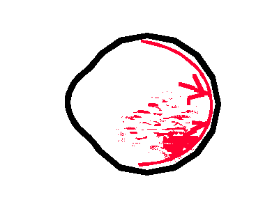
| This vitreous gel may start to shrink, and pull on the
growing new vessels, and may make them bleed. The bleeding usually causes a 'spiders web'
to appear in the vision, swirling around as the eye is moved. The blood is eventually
reabsorbed by the body's cells, and itself causes no damage. (But see below.) |
 |
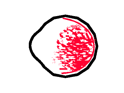
| A dense haemorrhage caused by a severe bleed still
usually clears itself, but problems may arise as below. |
 |
|
It is common to have such haemorrhages in proliferative retinopathy:
- if you have had a lot of laser, i.e. 6000 burns on
each eye, such bleeding is not usually serious, but more laser is usually needed.
- if you have had no laser the bleeding suggests that
quite a lot of laser may be needed, resonably soon (how soon: this depends on how severe
the condition is, perhaps within 2-4 weeks).
- if you have had perhaps 3000 burns, more laser is needed (how soon:
again this depends on how severe the condition is).
If you have a haemorrhage (it is impossible to give specific advice;
these are general principles)
- don't panic
- rest for a day or two sitting upright in a chair during the day, have
extra pillows to keep your head high at night
- let your eye clinic know (unless the bleeding is small, and
have had frequent haemorrhages, and have had 8000 or so burns on the eye,
and have had a recent examination)
- if your sight is badly affected it is safer to have your eye examined
by your ophthalmologist, although usually there is no immediate treatment. Laser is
usually arranged at a later date when it is likely much of the blood will have cleared.
|
Bleeding into the vitreous may contribute to the vitreous shrinkage.
If the shrinkage is mild the retina may
become slightly lifted or wrinkled.
Fortunately the wrinkling is usually away from the macula, and the sight should be good.
Surgery is not needed.
An 'epiretinal membrane' is the term given to one type of this condition, although it is
not always caused by bleeding. |
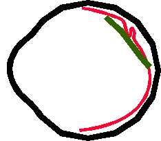
The retina (in red) becomes wrinkled
as the surface of the vitreous gel (in green) shrinks.
|
If this happens in the macular area (the
macula is described in 'mechanisms') your sight may be affected:
objects may appear tilted or bent. An operation (vitrectomy, as below, may be needed).
Shown here is a small 'traction detachment'. |
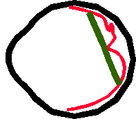
The vitreous shrinkage (green)
pulls on the central area of the retina (the macula) and affects the vision.
|
| If the condition is very severe, your
sight may be extremely bad. Vitrectomy surgery is usually helpful, but your sight may be
permanently damaged. |

The vitreous shrinkage is very severe,
pulling the retina (a large 'retinal detachment') |
If the vitreous shrinks and pulls the retina substantially, a
vitrectomy may be needed (as in the two paragraphs above).
| Similarly, if there is a dense
haemorrhage, surgery may be needed. An ultrasound test may tell the surgeon whether the
retina is in place or not (it can detach hidden behind the haemorrhage). This is a very
simple test using a scanning probe placed over your eye. A
vitrectomy carried out by an experienced surgeon is usually sucessful, but is not
discussed here in detail. The operation usually produces a cataract in the period after
the operation: this needs a cataract operation.
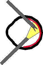
Three small holes are placed in the side of the eye, for instruments
like a special light, tiny scissors, and a vitreous 'cutter'. The blood is sucked out with
one of the probes, and if thickened membranes like those illustrated above are present,
they are peeled off the retina then sucked out. |
 |
This is a very nasty type of glaucoma that can occur in diabetes.
It occurs when when 'new vessels' grow and stop fluid draining out of the eye.
Treatment involves a lot of laser. (This explanation will hopefully be in more
detail in the next version of the sight.) See Glaucoma.
|