|
| |
Retinal vein occlusion
|
The retinal veins are the small ‘pipes’ in the retina that
drain blood out of the retina, back to the heart.
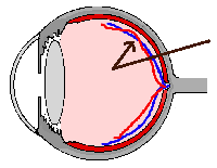 |
the veins of the retina may block
(veins in blue) this is a side
'cut through' diagram of an eye |
The retina is the thin film that lines the back of the eye, similar
to the film of a camera. It is the part of the eye that makes us see. Light (things that
you see) enters in through the front of the eye and falls on the retina.
The retina turns the light into electrical signals that are sent to
the brain, allowing you to see. (This is explained in more detail in HOW
RETINOPATHY DEVELOPS ).
The veins drain the blood out of the eye, whilst the retinal
arteries are the small pipes that deliver the blood (from the heart) to the retina, shown
in red in these diagrams. These arteries deliver the blood to the whole of the retina. |
| retinal vein
retinal artery |
|
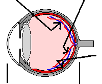 |
retina |
| front of eye
back |
|
| What is a retinal vein occlusion?
A retinal vein occlusion is a blockage of one of these veins.
The vein blocks when the blood in it stops flowing, and
the vein cannot then drain the blood out of the retina.
If the vein blocks some blood then leaks out of the vein. Also, clear fluid leaks out
causing ‘water-logging’ the retina. This naturally damages the sight.
|
|
Doctors believe that blood flowing through the
vein may be blocked by something pressing on the vein. For example, any condition, such as
high blood pressure for many years, that makes the small arteries ‘hard’, may
cause the artery to press on the vein and block it.
The distance between the arteries and veins varies in different
people, which may be one reason why some people are more likely to develop a blocked vein
than others. |
|
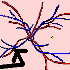
an artery-vein crossing point |
|
A view of the retina
from the front: what the doctor sees looking into your eye. The arrow points to an
artery—vein crossing point, where the vein may be blocked
|
| A blockage may be caused by something pressing on the
vein. For example, sometimes the small arteries can press on the vein: anything that makes
the small arteries ‘hard’ can lead to pressure on the vein. The distance between the arteries and veins varies in different
people. |
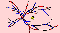
| The centre of the retina is responsible for your
sharp vision, such as seeing people’s faces or watching television. If this central part of the retina (a tiny yellow spot in
these diagrams) becomes ‘waterlogged’ by leakage from the blocked vein, your
sight will be reduced.
After approximately 3 months, if the waterlogging
remains, laser treatment may help to seal any leaks.
|
 |
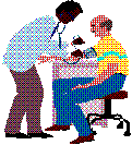
High blood pressure
|
Controlling the blood pressure
helps to prevent the arteries getting ‘harder’, and can prevent a blocked vein
in the other eye. |
|
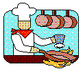
Too much fat in the blood
|
A healthy diet helps. The
Department of Health (& World Health Organisation) recommends people can help themselves by:
- five portions of vegetables or fruit every day
- 30 minutes exercise a day (eg walking, swimming)
- small amounts only of animal fat (meat, dairy food)
salt: too much may contribute to blood pressure
|
|
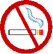
Smoking
|
this hardens all the arteries. The more you smoke, the more
damage is done (such as hardened arteries). Try to stop: ask your GP or nurse if you need
help. |
|
Glaucoma, diabetes, and other conditions
|
Other conditions may also cause a blockage of
a retinal vein, and extra treatment may be needed. Your doctor will check that you do not
have one of these conditions. |
|
Types of retinal vein occlusion
|
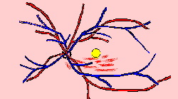
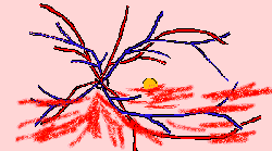
If the central area of retina, the macula (shown in yellow) is not
affected, the vision may be completely normal. The red marks show the 'heamorrhages' in
the retina.
|
 |
|
Inevitably the central part of the retina is
affected, reducing your sight, and laser is often needed. Laser treatment may be needed to
reduce waterlogging, stabilising the sight.
|
 |
|
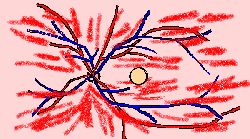
Unfortunately, your sight is usually affected in this type of
blockage. (Although a very mild blockage may not affect your sight.) Laser treatment does
not improve the sight, but it may be necessary to prevent complications: tiny blood
vessels can grow where they should not, leading to bleeding later.
If the blockage is severe, a lot of laser is
needed to prevent severe glaucoma.
|
 |
|
‘Laser’ is a very bright, but very narrow,
beam of light. You need to sit in a machine like the one used to examine your eye, and the
light is shone in through a small contact lens.
If your condition is mild, the laser treatment does not
usually hurt. |
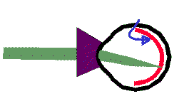
Laser light (green) is shone into the
eye through a small contact lens, and makes small burns on the retina (blue arrow).
LASER is simply a highly focused and
powerful light, where the light rays are all of the same type. For this reason it
can be pointed at one spot very accurately. |
- A healthy diet and regular exercise can help. See HELPING YOURSELF.
- Aspirin may help to prevent further blockages, and it
also helps to prevent heart disease. Ask your GP, especially if your have a peptic ulcer,
indigestion, or very high blood pressure.
- 'Statin' drugs may help, but many doctors are awaiting results of
reasearch. Ask your doctor.
- If you have glaucoma, the eye pressure still needs treatment. If you have
diabetes, you need to keep your sugar controlled and your blood pressure low. See PREVENTING PROBLEMS
- Generally patients you have had a retinal vein
occlusion need their eyes examining by an ophthalmologist for two years to determine
whether or not laser is needed. Patients with a minor blockage may not need many
examinations, and those with a severe blockage may need a longer follow up. Once
discharged from the hospital or clinic, see your optometrist every year for checks.
|
How to
cope with poor vision in one eye
Ask your eye clinic doctor and optometrist, and see COPING WITH POOR VISION and COPING WITH POOR
VISION IN ONE EYE.
|