|
| |
glaucoma
This is a condition that may occur in people with and
without diabetes.It is usually caused by too much fluid pressing on the nerve at the back
of the eye, as explained below in more detail.Parts of the
eye involved
|
 |
The eye is a round ball the size
of a small tomato. The arrow points to the optic nerve.
|
|
|
|
 |
It is partly hidden behind the
eyelids. |
|
|
|
 |
Imagine the eye turning as shown
below... |
|
|
turning more |
 |
|
and more |
 |
| then imagine 'cutting' through
the eye ball.... |
 |
|
 |
.....this
is the view used below to explain glaucoma.
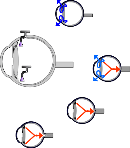
The blue arrow shows the flow
of fluid, like water, through the eye. |
The optic nerve in glaucoma
The optic nerve is the 'electric wire' of the eye, an it takes
messages about what you see on towards the brain. In the main type of glaucoma the optic
nerve is pressed on by extra fluid in the eye, and this may damage the sight in the eye.
How does glaucoma develop?
Everybody's eye produces a fluid like water in it's middle chamber.
This fluid then flows around inside the eye to the front chamber, as shown in the diagram
below... the blue arrow.
Then , from the front chamber the fluid leaves the eye by entering a drainage meshwork,
like the drainpipe of a sink or bath. From this drainage system the fluid enters the
bloodstream.
In the common type of glaucoma this drainage system can block. The
fluid gets trapped in the eye, and the pressure inside the eye goes up like a tyre being
blown up to much.
This pressure or fluid then presses on the nerve at the back of the eye. If the pressure
is high or continues for a long time, usually years, the nerve at the back of the eye may
become damaged, and eventually the sight may be affected. This pressure effect is shown by
the red arrow in the diagram below.
|

the optic nerve (red arrow) |
 |
The eye produces its own
fluid, like a tap in the bathroom', in its middle chamber.
The fluid ...like water.. flows
through from the middle to the front chamber, the eye, shown by the blue arrow. It then
comes out of the eye through a 'drain', like the plughole of a sink.
If the drain blocks, the fluid
cnnot get out of the eye, and the pressure in the eye builds up like a car tyre being
pumped up too much.
This pressure (red arrow)
damages the optic nerve at the back of the eye, pressing it in. |
The sight in glaucoma

At first the sight is normal, then a small area of poor vision may develop.
This can extend, affecting much of of the
sight,
or nearly all the sight.
A common type of loss of vision in
glaucoma. |
Severe glaucoma is not common if you have
diabetes, but it can occur. At first the sight is normal, but it if the glaucoma is
severe, the sight may get progressively worse as opposite.
You cannot 'feel' glaucoma, and usually would would not know there was anything wrong in
the early stages.The optometrist or ophthalmologist usualy
tests people with diabetes for glaucoma about once a year, as below.
|
How does the doctor or optometrist know you have
glaucoma?
Glaucoma is found by an ophthalmologist or optometrist by
- measuring the eye pressure
- looking into the eye at the optic nerve (the nerve can appear caved
in)
- testing the field of vision: you sit in a special machine with your
head still. Lights flash, and you are asked to press the button if you see the light. If
you cannot see the brighter lights, this shows on the computer print out rather like the
drawing of the 'castle' above.
Treatment for glaucoma
The basic treatment for glaucoma in diabetes is eye drops, and the
commonest is one of the beta-blocker drops such as betaxalol or timolol. These drops
switch the tap off that makes the fluid. You generally should not use these drops if you
get asthma or breathing difficulties, and should use an alternative drop. They may slow
the heart down (if they make you dizzy you should stop them) or make your ankles swell.
Will the sight get worse?
Once the pressure reaches a satisfactory level the glaucoma should not
get worse, or get very slowly worse.
A satisfactory pressure if the optic nerve is healthy is 18-20mmHg, but if the nerve is
already damaged the pressure needs to be lower, and 14 would be ideal. (The pressure level
needed also depends on your type of glaucoma. People with 'low tension glaucoma' need a
lower pressure, for instance.)
| Above one treatment has been
discussed. In the next revision of this site more details of other web-sites discussing
details of other drops and operations will be published. The explanation above relates to the usual type of glaucoma that may
occur in diabetes.
A very severe glaucoma is mentioned on this web-site (rubeotic glaucoma); this and other types
of glaucoma are discussed in more detail on other sites, although the basic mechanism is
similar to that above. |
See Royal National Institute for the Blind= http://www.rnib.org.uk
or the National Institiute of Health http://www.nei.nih.gov/
publications/glauc-pat.htm for more information about glaucoma, if this is your main
problem. |
|