|
| |
Retinopathy,
Mechanisms
The ‘retina’ is the film at the back of your eye, just
like the film in a camera.
Retinopathy is a disease of the retina, occurring in about a quarter of people with
diabetes.
How does the eye 'work'? Light enters the eye from the front, and passes through the eye
to hit the retina, just like in the camera. The retina contains cells that convert the
light into the electric signals, and these signals are then sent on to the brain so we can
see.
Two types of diagram are used in the descriptions in this section about retinopathy.
First, a side or 'cut through' view of the eye, like a cut through drawing of a camera as
opposite (upper picture).
Secondly, the view the doctor sees when he looks into your eye, like
a map, with the blood vessels spreading out from the centre (the optic nerve). This is
shown immediately opposite.
continued below.. how the retina works.. |
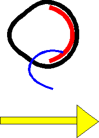
|
Light
enters the eye from the left in this diagram...shown by the yellow arrow.
It passes through the clear jelly of the eye (the vitreous) to reach the retina
(indicated with the blue pointer). |
| 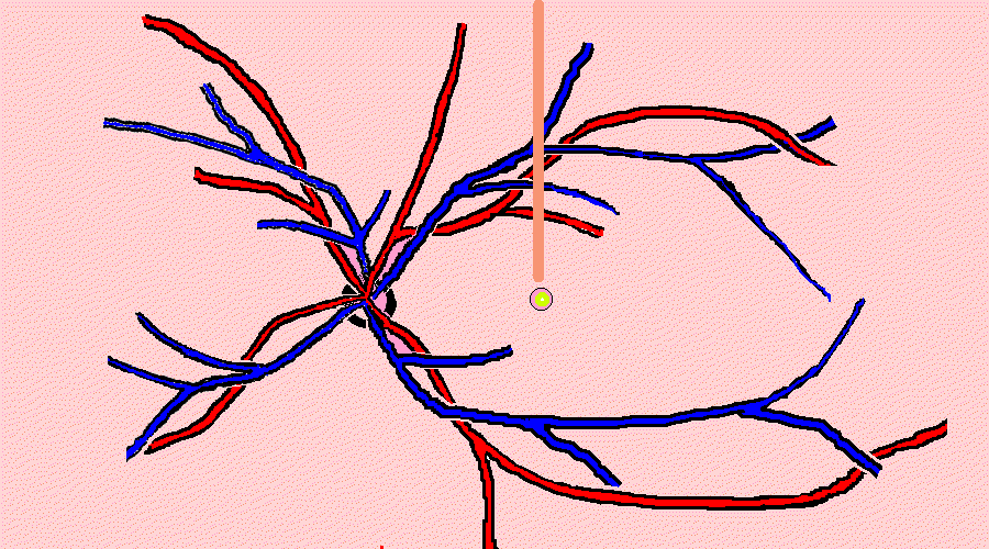
The retina: the view the doctor sees
looking into your eye (the pink marker points to the centre of the retina, where light is
focused).
The red & blue lines are the larger retinal blood vessels spreading out from
the optic nerve (black ring). |
|
How does the retina work?
The retinal cells stand next to each other,
a bit like houses in a street. The main cells are the rods and cones: these are the cells
that take up light and convert it into electrical messages, which are then sent onto the
brain.
These cells receive their oxygen and other nutrients from tiny blood vessels nearby. These
blood vessels are like pipes which pass nearby the cells; imagine a largish pipe passing
past your house, containing blood. The walls of these pipes/blood vessels are very thin,
and so nutrients can pass through them. These nutrients are the ‘food’ for the
cells. |
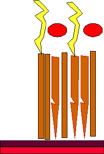 |
Light ...in yellow... falls onto the retina. The retinal cells are rods (the long
straight cells) and cones (the cells with the pointed end).
There are tiny blood vessels (capillaries) on the surface of the retina ...the red
ovals.
|
Retina in Diabetes (a simplified explanation)
If you are
diabetic you are likely to have a slightly high blood sugar. Over the years the high sugar
level can damage the tiny blood vessels.
The longer you are diabetic, especially if you have been diabetic for 14 years or more,
the more likely this is to happen. This damage can be slowed down by controlling your
sugar and blood pressure etc, and this is discussed in Preventing
Problems.There are three basic components of this
damaging process.
--the blood vessels can leak
--they can make a special growth substance that makes other vessels grow
--the vessels may eventually close and block.
First, the blood vessels can leak. This causes the retina to swell
up a little and become waterlogged, a bit like a sponge. This swelling then damages the
retinal cells themselves. This is the main mechanism in ‘maculopathy’.
This process is like a very leaky sieve or a raincoat that lets water in, instead of
keeping it out. |
|
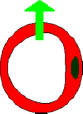
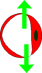
A healthy capillary, a tiny blood vessel . Nutrients pass out of the
capillary to reach the retinal cells, and waste products from the retina pass into it to
be taken away.
|
|
In diabetic retinopathy the damaged capillaries start to leak fluid.
|
| Secondly, the blood vessels can make a
special growth chemical that makes other tiny, tiny blood vessels grow. These are called
‘new’ blood vessels, and an ophthalmologists calls these ‘new
vessels’. These new vessels are very delicate and very easily bleed, and this blood
can (if the eye is not lasered) damage your eye badly. This is ‘proliferative
retinopathy’. |
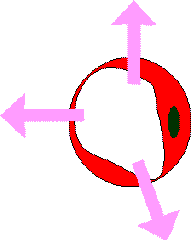 |
The damaged capillaries start to make a special growth chemical that makes
other capillaries grow.
|
| The tiny blood vessels may eventually close
and block. If the retina is badly damaged by leakage or very severe diabetes, the blood
vessels may close up, and nutrients will not reach the retinal cells. This happens in
‘ischaemic’ macular disease. |
The
capillaries start to close up and block. The retinal cells nearby can become damaged, and
the sight reduced.
|

|
These processes occur in the different types of diabetic retinopathy
as discussed on adjacent pages (see below for links).
|