EASDEC European Association for the Study of Diabetic Eye Complications

HOME    wpe19.gif (873 bytes)  CONTENTS   wpe18.gif (873 bytes)   SITE NEWS   wpe17.gif (873 bytes)   FEEDBACK    wpe16.gif (873 bytes)    HOT-LINKS   wpe14.gif (873 bytes)     EASDEC 99 MEETING


General pages

PATIENT INFORMATION

CATARACTS

WEB-SITE NEWS

COPING WITH POOR VISION
& RELATED PAGES

PREVENTING PROBLEMS

RETINOPATHY MECHANISMS

TYPES OF RETINOPATHY

BACKGROUND RETINOPATHY

MACULOPATHY

PRE-PROLIFERATIVE

PROLIFERATIVE

GLAUCOMA

SORE EYES

VITREOUS HAEMORRHAGES, EPIRETINAL MEMBRANES,
TRACTION RETINAL DETACHMENTS, VITRECTOMY, RUBEOTIC GLAUCOMA

LASER TREATMENT

RETINAL VEIN OCCLUSION


Health Professional

ABSTRACTS & RESEARCH

SCREENING

LOW VISION & REHABILITATION

EASDEC MEMBERSHIP

CONTRIBUTIONS TO THIS SITE

LIST OF MEMBERS

EASDEC 99 MEETING

JOURNAL REVIEW


Background retinopathy

Your sight should be perfectly good at this stage. A doctor examining your eye will notice tiny abnormalities.
Your pupils have to be dilated  for this examination, and you are often advised not to drive until the pupils have returned to their normal size.
The tiny blood vessels in the retina, the capillaries, become damaged. This has been discussed on the 'Mechanisms of Retinopathy' page.

What the doctor sees

A doctor or optometrist may see 'dots' and 'blots'.

The dots are some capillaries that have enlarged, that is the the tiny blood vessels enlarge to form microaneurysms.

The blots are tiny haemorrhages, that is tiny spots of blood, on the surface of the retina.

What does it mean if you have 'background retinopathy'?

The number of microaneurysms, the little red dots the doctor sees, indicate the likelihood of more severe problems in the years to come.

Retinopathy develops more slowly if your sugar and blood pressure etc (PREVENTING PROBLEMS) are controlled. Unfortunately for many people with diabetes the retinal damage increases, and maculopathy or proliferative retinopathy develop over a few years.

wpe21.gif (3361 bytes)

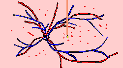
This is the view a doctor sees looking into your eye. The small red dots are 'microaneurysms', tiny damaged capillaries. The red lines are small haemorrhages, little flecks of blood. Your sight is not affected at this stage.


Home ] Up ]

Send mail to [email protected] with questions or comments about this web site.
Last modified: April 06, 1999
About Easdec
EASDECs 1999
         meeting

Easdec: joining
Easdec list of
        members

Abstracts x 11
Feedback
Patient Information
Visual Aids etc
Coping..one eye
Coping with poor vision:4 pages
Vitreous haemorrhages, traction
        detachments, etc
Mechanisms of Retinopathy
Types of retinopathy
Background
Maculopathy
Pre-proliferative
Proliferative
Contents
hot links
Site News
Laser
Sore/dry eyes
Glaucoma
Journal Review
Contributions
Cataracts
Preventing Problems
Screening
Screening,
      comprehensive
       review

diabetes.uk web site= http://www.diabetic.org.uk/main1.htm
British Diabetic Association= http://www.diabetes.org.uk
Royal National Institute for the Blind= http://www.rnib.org.uk/info/eyeimpoi/diabetic.htm